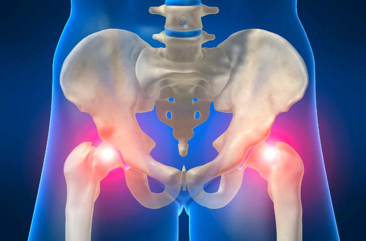
CAM and Pincer Impingement Decompression
An arthroscopic procedure performed to remove bony impinging spurs from the acetabulum (socket) and femoral head (ball). Two or three small incisions are made around the hip. An arthroscopic camera and instruments are used to identify the impinging structures. A burr is used to remove excessive bone. Hip range of motion is examined under live xray during the surgery.

Common Questions About CAM and Pincer Decompression
What is CAM and Pincer impingement?
CAM impingement of the hip is when the femoral head, or the ball of the femur is aspherically shaped and does not fit perfectly inside the acetabulum, or the socket of the hip. This condition can lead to pain and early onset loss of cartilage leading to early arthritis.
Pincer impingement is when the acetabulum or socket of the hip, displays overcoverage of the femoral head of the femur which results in the constant pinching of the labrum which can lead to labral tears.
What is the surgical procedure like?
During impingement decompression of the hip, the surgeon makes 2-3 small incisions on the hip and a burr is used to remove bony protrusions on either the femoral head or acetabulum.
What is the recovery process like?
Following the surgery, the patient will use crutches for the first few weeks and then transition to weight bearing activities. Full recovery from the surgery can take from 4-6 months. Patients start attending physical therapy when instructed by their physician and continue for the duration of their recovery process.


